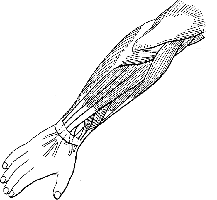
Diagram Of The Muscles In The Forearm / Print Muscles flashcards | Easy Notecards / Because the contribution of each forearm muscle to elbow movement is small, it is often not recognised in conventional anatomy teaching.
Diagram Of The Muscles In The Forearm / Print Muscles flashcards | Easy Notecards / Because the contribution of each forearm muscle to elbow movement is small, it is often not recognised in conventional anatomy teaching.. A deep layer , intermediate layer and superficial layer. Start studying muscles of the forearm. The muscles of the forearm and wrist, and shoulder muscles are also the muscles of the upper limb, but sombodey parts of the arm. 2, ulna, 3, biceps muscle; Arm muscle diagram, forearm front arm muscle anatomy muscle diagram arm anatomy, anatomy of shoulder ligament ideas anatomy lesson full hd from the arm muscle diagram above, the muscles of the arm that can be seen easily on the surface include biceps, triceps, brachioradialis, extensor.
Related posts of muscles of the arm and forearm diagram. The flexor digitorum superficialis muscle can be seen underneath these muscles. The term forearm is used in anatomy to distinguish it from the arm. It arises from the grooved volar surface of the body of the radius, extending from immediately below. Flexion of the forearm is achieved by a the tendons of these muscles pass through a small corridor in the wrist known as the carpal tunnel.
Label and Color the Muscles of the Arm (Extensors) from www.biologycorner.com Muscles allow a person to move skeletal muscles are the only muscles that can be consciously controlled. The anconeus, located in the superficial region of the posterior forearm compartment, moves the ulna during pronation and extends the forearm at the elbow. Learn vocabulary, terms and more with flashcards, games and other study tools. Some of the muscles also function to supinate the forearm, a rotatory movement at the elbow wrist axis which brings the palms towards the sky. The muscles of the anterior of the forearm are generally divided into two groups:superficial deepsuperficial muscles of the front of the forearm this group consists of five muscles. It is a functionally important muscle that contains two heads. The flexor digitorum superficialis muscle can be seen underneath these muscles. The anterior forearm muscles are divided into 3 muscular layers ;
Related posts of muscles of the arm and forearm diagram.
Try labeling diagrams and worksheets as additional learning aids. The antibrachial or forearm muscles may be divided into a volar and a dorsal group. This is the most medial of the superficial flexor muscles in the forearm. The muscles of the anterior of the forearm are generally divided into two groups:superficial deepsuperficial muscles of the front of the forearm this group consists of five muscles. The forearm is the region of the upper limb between the elbow and the wrist. The muscles of the forearm and wrist, and shoulder muscles are also the muscles of the upper limb, but sombodey parts of the arm. Learn vocabulary, terms and more with flashcards, games and other study tools. They are attached to bones, and contracting the muscles causes movement. The anterior forearm muscles are divided into 3 muscular layers ; Diagram the movements of the humerus muscles that act on the forearm. Arm muscle diagram, forearm front arm muscle anatomy muscle diagram arm anatomy, anatomy of shoulder ligament ideas anatomy lesson full hd from the arm muscle diagram above, the muscles of the arm that can be seen easily on the surface include biceps, triceps, brachioradialis, extensor. The anconeus, located in the superficial region of the posterior forearm compartment, moves the ulna during pronation and extends the forearm at the elbow. Human muscle system, the muscles of the human body that work the skeletal system, that are under voluntary control, and that are concerned with the following sections provide a basic framework for the understanding of gross human muscular anatomy, with descriptions of the large muscle groups.
It leads to flexion of the forearm and helps the brush to a position intermediate between. The term forearm is used in anatomy to distinguish it from the arm. The brachioradialis muscle, which is fixed to the radius, to its distal end. Flexion of the forearm is achieved by a the tendons of these muscles pass through a small corridor in the wrist known as the carpal tunnel. Learn vocabulary, terms and more with flashcards, games and other study tools.
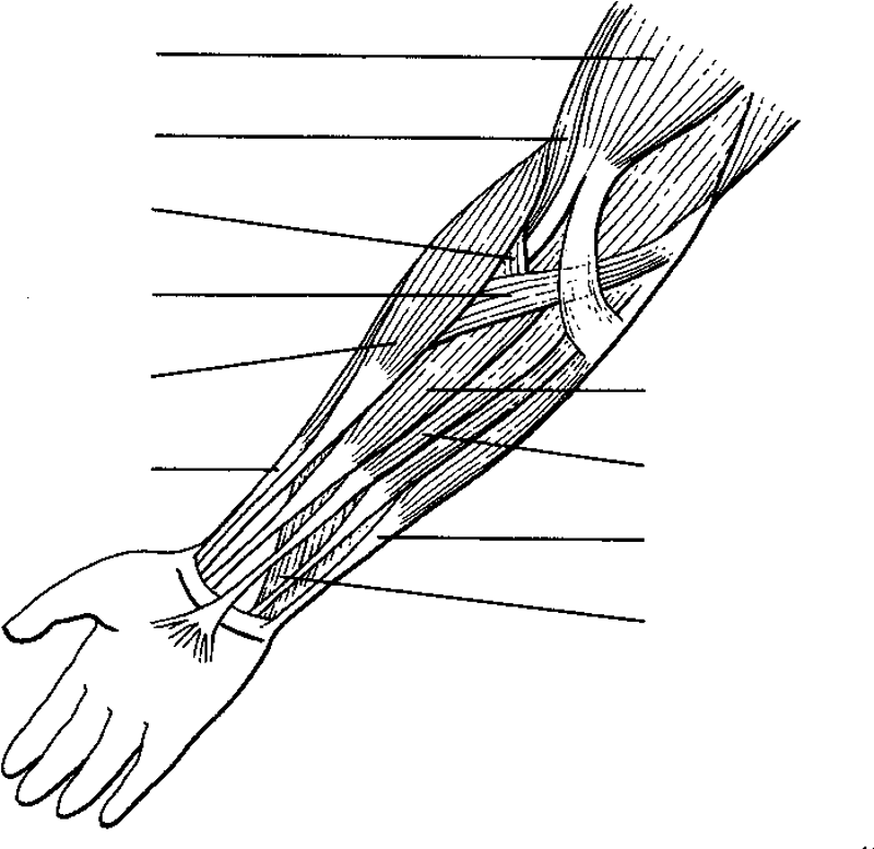
Muscles of the Forearm | AnatomyZone from anatomyzone.com Diagram the movements of the humerus muscles that act on the forearm. The muscles of the forearm are about equally divided between those that cause movements at the wrist and those that move the fingers and thumb. The forearm is the region of the upper limb between the elbow and the wrist. Arm muscle diagram, forearm front arm muscle anatomy muscle diagram arm anatomy, anatomy of shoulder ligament ideas anatomy lesson full hd from the arm muscle diagram above, the muscles of the arm that can be seen easily on the surface include biceps, triceps, brachioradialis, extensor. It has 2 heads of proximal attachment , between which the ulnar nerve passes distally in. Learn vocabulary, terms and more with flashcards, games and other study tools. Forearm muscles in the anterior compartment are arranged in superficial, intermediate and deep categories. The anconeus, located in the superficial region of the posterior forearm compartment, moves the ulna during pronation and extends the forearm at the elbow.
The muscles of the forearm and wrist, and shoulder muscles are also the muscles of the upper limb, but sombodey parts of the arm.
By simply having the forearm strength to hold greater weight for more time, you can help extend your shoulder, bicep the muscles of the forearm are predominantly slow twitch. The main muscles of the forearm can make or break a fantastic workout and physical routine, so here you will get some of my favorite exercises to strengthen the forearm muscles along with some hidden advantages to become large forearms. Muscle anatomy diagram 12 photos of the muscle anatomy diagram canine muscle anatomy diagram, dog muscle anatomy diagram, lower leg muscle anatomy diagram, muscle anatomy of human back, tricep muscle. Start studying muscles of the forearm. Inflammation of this region caused by repetitive. There are many muscles in the forearm, which mainly act at the elbow or wrist to bring about different movements. The forearm is the region of the upper limb between the elbow and the wrist. Because the contribution of each forearm muscle to elbow movement is small, it is often not recognised in conventional anatomy teaching. 4, attachment… the muscles of the back forearm. The muscles of the upper arm are responsible for the flexion and extension of the forearm at the elbow joint. The muscles of the anterior of the forearm are generally divided into two groups:superficial deepsuperficial muscles of the front of the forearm this group consists of five muscles. Superficial muscles of the posterior forearm: It arises from the grooved volar surface of the body of the radius, extending from immediately below.
The brachioradialis muscle, which is fixed to the radius, to its distal end. The term forearm is used in anatomy to distinguish it from the arm. 2, ulna, 3, biceps muscle; The forearm is the region of the upper limb between the elbow and the wrist. The muscular system consists of various types of muscle that each play a crucial role in the function of the body.
Print Muscles flashcards | Easy Notecards from www.easynotecards.com The flexor pollicis longus is situated on the radial side of the forearm, lying in the same plane as the preceding. Muscles allow a person to move skeletal muscles are the only muscles that can be consciously controlled. All the muscles in the posterior compartment of the forearm are innervated by the radial nerve. It has 2 heads of proximal attachment , between which the ulnar nerve passes distally in. The muscles of the upper arm are responsible for the flexion and extension of the forearm at the elbow joint. Muscle anatomy diagram 12 photos of the muscle anatomy diagram canine muscle anatomy diagram, dog muscle anatomy diagram, lower leg muscle anatomy diagram, muscle anatomy of human back, tricep muscle. Start studying muscles of the forearm. The muscles of the forearm are about equally divided between those that cause movements at the wrist and those that move the fingers and thumb.
Start studying muscles of the forearm.
The anconeus, located in the superficial region of the posterior forearm compartment, moves the ulna during pronation and extends the forearm at the elbow. This layer contains only one muscle, the flexor digitorum. The superficial extensors of the forearm are the brachioradialis, extensor carpi radialis longus, anconeus, extensor carpi radialis brevis, extensor carpi ulnaris, extensor digitorum and extensor digiti minimi. The main muscles of the forearm can make or break a fantastic workout and physical routine, so here you will get some of my favorite exercises to strengthen the forearm muscles along with some hidden advantages to become large forearms. The muscles of the upper arm are responsible for the flexion and extension of the forearm at the elbow joint. Editor · aug 11, 2017 ·. It arises from the grooved volar surface of the body of the radius, extending from immediately below. 2, ulna, 3, biceps muscle; Learn vocabulary, terms and more with flashcards, games and other study tools. The forearm is the region of the upper limb between the elbow and the wrist. The anterior forearm muscles are divided into 3 muscular layers ; Start studying muscles of the forearm. A deep layer , intermediate layer and superficial layer.


0 Komentar